G-Files
New rotary NiTi glide path instrumentation! |
| |
Glide path development is an essential but time-consuming step in endodontic treatment. To increase endodontic efficiency in the initial glide path formation by simplifying the procedure while increasing safety, MICRO-MEGA ® is introducing two new rotary NiTi instruments, the G-Files. The G-File instruments are based on an innovative design to help the clinician safely save time in endodontic procedures. The superior cross-section of the G-Files combines efficiency and innovation. The G-File NiTi instruments are machined with a narrow diameter (12 and 17) with a slight .03 taper. The superior cross-section of the G-File combines efficiency and safety. Along the length of the instrument, the G-File has cutting edges on three different radiuses, creating a large and efficient area for upward debris removal.
|
| |
|
| |
The angular offset of the cutting edges also creates a different pitch along the length of the blade, avoiding any screwing or engaging effect into the walls of the canal. The non-working (safety) tip is asymmetrical which helps the instrument safely move forward; this is also facilitated by the high degree of flexibility resulting from the small diameter.
The G-Files are electro-polished, which improves their mechanical properties, particularly by releasing internal stresses which develop during machining, thereby increasing the flexibility of the G-File. The electro-polished surface increases the efficiency in apical progression of the G-File while aiding in debris removal. The G-Files are available in 21, 25 and 29 mm.
Used after hand files have measured working length, G-Files safely enlarge the glide path in preparation for RCT with rotary instrumentation system. |
| |
---------------------------------------------------------------------------------------------------------------------------------- |
| |
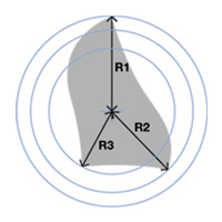
| Unique innovative cross-section |
| |
 |
|
- |
The cross-section varies throughout the length of the instrument. |
- |
The 3 cutting edges are on 3 different radiuses relative to the axis of the canal. |
- |
More space for better elimination of debris. |
- |
Excellent cutting action. |
|
| |
|
|
|
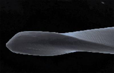
Non-working Tip |
| |
|
| |
SEM view: Dr Franck Diemer,
Toulouse, France |
|
---------------------------------------------------------------------------------------------------------------------------------- |
| |
Protocol for use: |
| |
|
|
| |
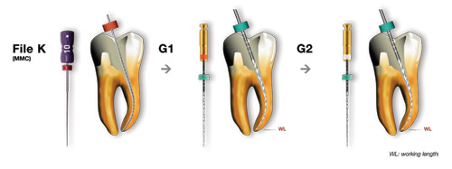
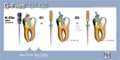
1. |
|
Determine the working length with a small diameter precurved stainless-steel instrument (MMC 08 and 10 files). |
2. |
|
The rotating G1 instrument is introduced into the canal, progressing with a slow movement without any apical pressure until the working length has been reached. |
|
3. |
|
After irrigation, the G2 instrument is used in the same way; then the last hand file is udes again to check patency and confirm the working length. |
|
|
|
| |
| Speed of rotation: 400 rpm – Max. torque: 1.2 N.cm |
| Note: It may be necessary to use ENDOFLARE to allow easy direct access of the G-Files to the entrance of the canal. |
| |
---------------------------------------------------------------------------------------------------------------------------------- |
| |
| Packaging/References: |
| |
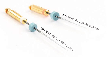
Two instruments are offered in steril and non-steril:
G1: N°12 - .03 - L21, 25 or 29 mm
Pack of four identical or assorted instruments (G1 x 2 + G2 x 2)
G2: N°17 - .03 - L21, 25 or 29 mm
Pack of four identical or assorted instruments (G1 x 2 + G2 x 2)
The G-Files exist in a Classics and InGet ® version. |
|
---------------------------------------------------------------------------------------------------------------------------------- |
| |
| Clinical cases: |
| |
 |
|
|
| |
|
|
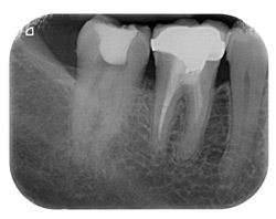
Pre-operative x-ray pulp chamber open for diagnosis of pulp necrosis |
|
First penetration with hard file MMC 08 to the WL (with apex locator)
|
| |
|
|
 |
|
 |
| |
|
|
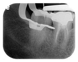
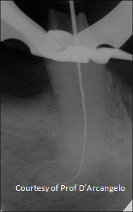
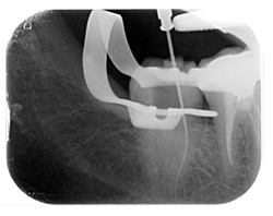
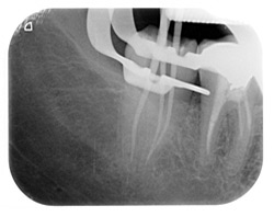
X-ray with G1 (electronic WL confirmed) |
|
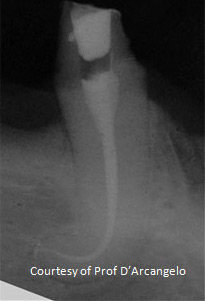
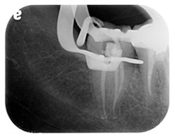
Radio G2 in place |
| |
|
|
 |
|
 |
| |
|
|
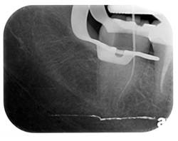
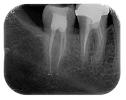
X-ray with gutta percha cones after the use of the Revo-S sequence : SC1-SC2-SU
(to the WL) and AS 30 to the WL 1mm |
|
Obturation with warm vertical condensation technique |
| |
|
|
 |
|
|
| |
|
|
| |
|
|
| |
|
|
|
|
|
| |
|
|
Fig. 1: Canal exploration and working length of the 3.5 (LL5) |
|
Fig. 2 Post operative x-ray after having used G-Files to obtain a right glide path, Revo-S to shape the root canal and HEROfill for canal filling.
|
| |
|
|
|
---------------------------------------------------------------------------------------------------------------------------------- |
| |
| Documentations : |
| |
|
| |
| ---------------------------------------------------------------------------------------------------------------------------------- |
|